Chanjong Park(통합과정, 제1저자)
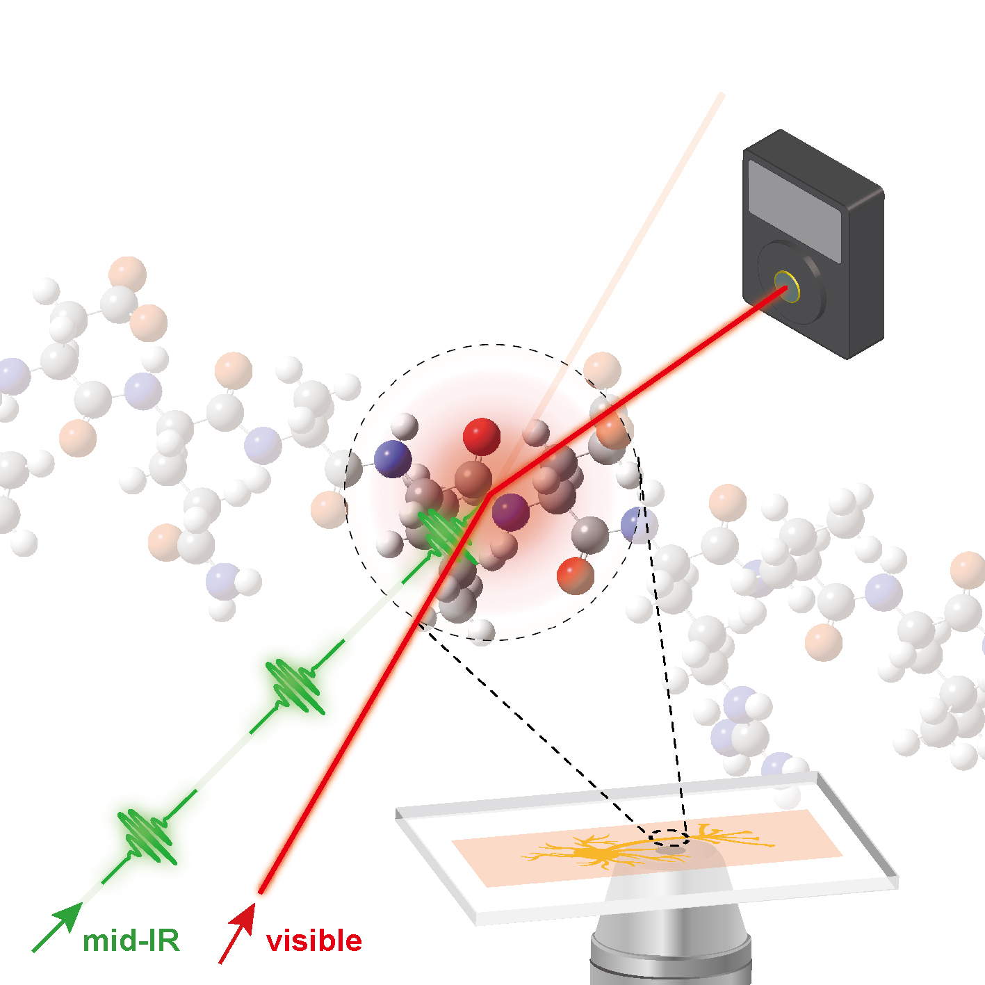
Mid-infrared photothermal microscopy has been suggested as an alternative to conventional infrared microscopy because in addition to the inherent chemical contrast available upon vibrational excitation, it can feasibly achieve spatial resolution at the submicrometer level. Furthermore, it has substantial potential for real-time bioimaging for the observation of cellular dynamics without photodamage or photobleaching of fluorescent labels. We performed real-time imaging of oligodendrocytes to investigate cellular dynamics throughout the life cycle of a cell, revealing details of cell division and apoptosis, as well as cellular migration. In the case of live neurons, we observed a photothermal contrast associated with traveling protein complexes on an axon, which correspond to the transport of vesicles from the cell body to the dendritic branches of the neuron through the cytoskeleton. We anticipate that mid-infrared photothermal imaging will be of great use for gaining insights into the field of biophysical science, especially with regard to cellular dynamics and functions.